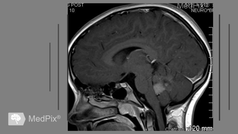
The tissue of medulloblastoma with extensive nodularity, which resembles the structure of a grapevine, has both malignant and benign cells. Scientists from Heidelberg were able to identify the mechanism of their transformation. This will help in the search for new methods of treatment for the most common brain cancer in children.
About the tumor
Medulloblastoma is one of the most common solid tumors and the most common childhood malignant brain tumor.

There are several histologic subtypes of this neoplasm, which can have very different courses. While some progress aggressively and form metastases, there are other forms that can usually be cured with intensive combination therapy including surgery, chemotherapy, and radiation.
One of those subtypes is medulloblastoma with extensive nodularity (MBEN). It is characterized by good recovery chances and special structural properties. Its tissue consists of many areas with nodules adjoining each other like grapes in a vine.
About the study
A group of scientists from the Hopp Children's Cancer Center, the German Cancer Research Center and the Heidelberg University Hospital set themselves the goal of finding out which factors at the cellular level direct the development of certain forms of medulloblastoma in a benign or malignant direction.
The object aforementioned medulloblastoma with pronounced nodularity became the study object. Using an innovative bioinformatic method, they were able to create a laboratory model of this type of tumor. This made it possible to determine that it contains both cells that develop very similarly to normal brain cells and those that have already become malignant. It all depended on where exactly they were located.
And in the intermediate zones, in addition to immune and connective tissue cells, significantly more aggressive tumor cells were detected, which continued to divide uncontrollably, and their genetic program was more similar to fast-growing medulloblastomas and immature nerve cells. However, in the process of migrating to the nodes, these cancer cells transformed back into normal nerve cells and stopped dividing.
Scientists' conclusions
Christian Peitler, pediatric oncologist and leader of the research project, explains the findings this way:
The scientist suggests that in the case of MBEN tumors, this works only partially, and many cells go through half of the normal cerebellar cell development process and stop dividing. This also explains the predominantly favorable course of this type of tumor.
Clinical relevance of findings
Blocking the maturation process is a key mechanism whose management will prevent malignant progression in children and guide cancer cell development in a benign direction.
He emphasizes that even if most children with this type of medulloblastoma can be cured with surgery and, if necessary, other therapies, these are very stressful and risky methods for young children, often associated with severe, lifelong side effects. The work of the Heidelberg researchers sets the stage for the possibility that in the future young patients could be treated in much safer ways.
References
- Ghasemi D.R., Okonechnikov K. et al. Compartments in medulloblastoma with extensive nodularity are connected through differentiation along the granular precursor lineage. In: Nature Communications (Online Publikation 8. Januar, DOI: 10.1038/s41467-023-44117-x)

Comments — 0