
Computed tomography is used to assess the condition of musculoskeletal structures and soft tissues. The procedure does not cause unpleasant feelings, provides a large amount of important information, and the amount of radiation exposure allows performing the examination as many times as necessary without harming the body.
Principles of CT technology
The method is based on the ability of tissues to absorb X-rays with varying degrees of intensity. As a result, cross-sectional images (slices) are obtained. High picture resolution is achieved by using a sensitive matrix and sensors. The received signal is converted and digitized.
A modern tomograph is a complex device with mechanical and electronic components. Sophisticated detector arrays sense X-ray radiation. They are being constantly upgraded, so error risks in findings obtained by state-of-the-art computed tomography scanners are reduced to zero. A key element is software which makes it possible to conduct the study with certain parameters, transform the image and analyze the data.
It is advisable to perform follow-up scans at the same facility and using the same system as the initial one in order to avoid possible discrepancies in findings.
What are the existing computed tomography types?
CT scan variations are classified according to various parameters, such as the scanning speed, application area, and the use of a contrast agent. The most important is the method of exposure.
Spiral CT
In this case, the beam of rays moves in a spiral, and with each rotation one or more images are recorded. The speed of the X-ray tube movement is set by the operator. In this case, the body is not exposed to a high radiation load during the procedure. The probability of error is 2%.
MSCT
Multislice computed tomography is a technique which enables obtaining images of several slices at once in one revolution of the tube. Depending on the system type, the number of slices can be up to 320.
The main advantages of MSCT are:
- the ability to detect the smallest abnormal changes thanks to the minimal slice thickness;
- short duration of the procedure (if a contrast agent is not used, the procedure takes half a minute);
- high image quality;
- wide area of application and possibility of combining with other diagnostic methods;
- possibility of saving the results in different formats for easy transfer between clinics;
- low radiation dose in comparison with conventional CT;
- application in complex bone and joint conditions;
- ability to adapt radiation load depending the examination area and individual characteristics of the patient.
Electron beam computed tomography (EBCT)
EBCT is one of the most novel examination tools. Its difference is in the use of four sector targets to obtain a large number of slices. They are stationary. At the same time, a beam of electrons from the gun moves in a circle. This allows to perform image sweeps in as little as 0.025 seconds. As a result, the tool can be used to scan the heart in stillness, since the pulsations of the organ are not picked up due to the ultra-short exposures. EBCT makes it possible to detect calcinates in the vessels, and correctly evaluate the heart condition in crying or moving infants.
Dual-source CT (DSCT)
The first device with two radiation sources was released in 2005. The aim was to scan the heart in constant motion. In DSCT this is achieved by synchronizing it with ECG. Hereby it is not possible to have the shortest time interval between the slices, which would allow to remove errors resulting from organ tissue movement.
However, if two tubes are placed at an angle of 90 degrees, the time of full rotation can be shortened and the resulting slice image will show the non-contracting heart. In addition, the tubes can work each in its own mode, enabling the detection of foci located close to the bones, or metal elements. This mode is used when the CT is performed with a contrast agent.
CT devices with two radiation sources have the same advantages as MSCTs.
Cone Beam CT
This method is used especially in dentistry and maxillofacial surgery. It allows you to identify bone abnormalities and create a three-dimensional model based on the results of the study. This feature is useful in bite correction, management of trauma and other disorders. The doctor uses the image to assess the condition of the bone tissue and make a treatment plan. In addition, the technique is also applicable in follow-up care to assess the treatment and rehabilitation effect.
Combination with other examinations
The scope of computed tomography can be enhanced by combining it with other diagnostic tools, such as positron emission tomography. PET/CT refers to hybrid modalities widely used, in particular for many cancers. The patient is injected with a radioactive drug, the natural decay of which produces low radiation, allowing to visualize tracer distribution in the body. In this case, the increased metabolism indicates the presence of abnormal, malignant changes. With a computed tomography (CT) scan performed at the same time, it is possible to determine exactly in which parts of the body this occurs. Thus, a CT scan combined with a PET scan serves to identify foci of cancer or its metastases, as well as to determine how far it has spread in the body.
Scope of computed tomography
The examination enables a millimeter-precise scan of internal body structures, creating the most accurate three-dimensional model. The method is highly informative in evaluating bony structures, as well as fluid-containing masses. General indications for CT scanning include:
- hemorrhages in the gastrointestinal tract;
- trauma of soft tissues;
- benign and malignant neoplasms;
- metastases and affected lymph nodes;
- abdominal, thoracic, and soft tissue abscesses;
- respiratory system disorders;
- infections;
- muscular dystrophy;
- acute circulatory disorder consequences;
- blood clots in vessels;
- congenital abnormalities in the structure and position of organs and systems.
Computed tomography: arguments in favour
CT examination is one of the standard methods of diagnostic imaging. An important advantage is the accuracy with which anatomical structures and pathologies are visualized. But there are other pros as well:
Speed: CT scans usually take only a few minutes. This is why it is the preferred method of examination in emergency medicine, for example, in the diagnosis of strokes.
Combinability: this technique can be very well combined with others to solve specific issues. The best example is the combination of positron emission tomography and computed tomography.
Reliability: CT scans can also be performed in the presence of a pacemaker or implants. This makes this method suitable for a wider target group than MRI.
No overlap effect: unlike conventional X-rays, CT scans do not provide a two-dimensional image in which overlap is possible. The images are taken from several angles, allowing structures lying one behind the other to be clearly seen.
How do I prepare for a CT scan?
There is practically no preparation required for the examination. In the case of pelvic, digestive and liver examinations, diet and colon cleansing may be necessary to avoid misrepresentation. If a contrast agent is to be used, e.g., examining the brain, a lab test is necessary to rule out an allergic reaction.
CT contraindications
The technique does not involve any organ or tissue damage. The X-rays passing through the tissues do not stir any abnormal processes. However, a CT scan should not be performed during pregnancy, because it involves a certain amount of radiation.
Contraindications to CT with contrast are broader due to the use of a tracer. It should be avoided in the following cases:
- thyroid disorders;
- allergic reactions to contrast agents;
- chronic kidney conditions with function disorders;
- severe form of diabetes mellitus.
Besides, the majority of CT devices have patient wight limitations, so the study can not be performed in the case of severe obesity.
What is the procedure?
The duration of a CT scan varies from one minute to half an hour, depending on the area to be examined and the use of contrast. The procedure is painless, but some discomfort may be caused by the sounds produced by the device.
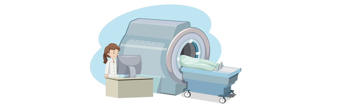
The patient is laid on the scanner table and is secured with straps and rollers to ensure immobility. After this, the table moves inside the machine while the findings are being obtained. Once the scan has been done, the table moves out. Processing the results may take some time, depending on the radiologist's workload.
Evaluation of results
The outcome of the procedure includes a written report, printed images and a CD recording. The CT report specifies the position of the organ, its borders, size, structures, and other details. If there are any abnormalities, the foci are described.
The reliability of these findings depends on the following factors:
- whether the patient was completely motionless during the examination;
- the presence of metal elements (implants, plates) in the body;
- device functioning;
- doctor's qualification.
Computed tomography enables identifying abnormal foci in the early stages, but it is only possible if the imaging is interpreted correctly. In complicated cases, it is recommended to seek more than one assessment, and the best solution is consulting European experts. A second opinion is a chance to get advice from a physician with a certain subspecialty, that is, expertise in the particular area to which your particular issue is related. The option is available online or in the written form; no personal visits are required.
References:
1. Computed Tomography: A Technical Review, Euclid Seeram, 2018.
2. Advances in computed tomography, Ellen C Benya, 2008.
3. Volumetric computed tomography: Principles, parameters, A Jankowski, 2010.
Comments — 0