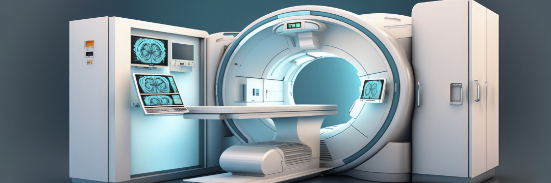
Assessing the nature, localization and extent of a lesion is of paramount importance in lung diagnosis. These questions are the domain of radiology. X-rays and CT scans are commonly used, but in recent years physicians have become increasingly aware of the diagnostic potential of MRI of the lungs and bronchi.
What is the study object?
Our lungs are vital organs. They enable us to breathe and supply oxygen to the whole body. When we breathe in, the air first enters the windpipe, which divides into two main bronchi - branches of the windpipe that enter the right and left lung hilus.
They continue to divide into thinner and thinner bronchioles, at the end of which are the alveoli. This is where the final gas exchange takes place: oxygen enters the blood and carbon dioxide is released from the blood into the air, which is then exhaled.
These range from pneumonia and bronchial asthma to cancer. In some cases, depending on the clinical picture and the individual circumstances, a chest MRI may be indicated to diagnose the conditions.
What is magnetic resonance imaging of the lungs and bronchi?
Magnetic resonance imaging provides images of these organs without using X-rays.
The lungs are scanned layer by layer, producing multiple two-dimensional cross-sectional pictures of high accuracy and resolution. These are then superimposed by a computer. The result is a three-dimensional image of the inside of the chest.
This method of imaging differs from others in the high contrast of soft tissues, which makes it easy to see even the smallest changes. The high-precision technique is therefore used to diagnose many diseases of the chest organs. It is also non-invasive, completely painless and has no risk of radiation exposure.
Lung MRI: a «young» technique with many applications
In comparison to CT and X-ray, the use of magnetic resonance in pulmonology is a relative novelty, as it was introduced into clinical practice later. As a result, the former two conventional methods are the most widely used. In addition, the technique has a number of technical challenges, such as signal loss due to cardiac and respiratory pulsation and artefact occurrence due to the presence of air-tissue boundaries.
Advantages
However, recent studies have shown that it can be as accurate as a CT scan, but without the radiation exposure. In addition, MRI is the right choice for children and pregnant women, for whom even the slightest exposure to radiation is contraindicated.
This means that both morphological changes and malfunctions can be assessed, including perfusion, ventilation, respiratory mechanics, cardiac function and blood flow.
Indications
The ability to combine different diagnostic aspects in one procedure makes this test useful in a wide variety of conditions, such as:
- pneumonia (lung infections);
- obstructive airway diseases (e.g., cystic fibrosis, bronchial asthma, COPD);
- pulmonary vascular diagnosis;
- lung cancer;
- pulmonary metastases;
- pulmonary embolism.
In obstructive airway disease, especially in cystic fibrosis, MRI is increasingly replacing CT as a procedure for diagnosing complications or monitoring the disease course.
How does magnetic resonance imaging represent tissues?
In general, tissues and organs containing water, such as liver or muscle, appear brighter, while areas with little water, such as the lungs, appear darker. Inflammatory processes, such as pneumonia, also look lighter than surrounding tissues.
Obstructive disease-specific changes (bronchiectasis, wall thickening, mucosal obstruction, and infiltrates) are very well seen, allowing for accurate clinical conclusions. The radiation-free diagnostic option is particularly important for patients with cystic fibrosis because of their potentially high cumulative lifetime exposure to radiation.
Until now, CT scans have been used for early detection, but studies show that MRI can be just as accurate and safe. In addition, because of the higher contrast of soft tissues, interrelation with surrounding structures can be better assessed with the help of the current techniques.
Lung MRI with contrast
In pulmonary vessel examination a contrast agent is often injected to ensure better imaging. The contrast makes them appear on the scans brighter than the surrounding tissues. Blood flow obstacles, such as emboli, also become clearly visible.
In addition, the contrast uptake intensity varies from tissue to tissue. This feature is also used in detecting tumors or metastases.
Helium as a contrast agent
A technique allowing the use of magnetic resonance to image lung ventilation has been under development for a few years. Before the examination, the patient inhales the harmless inert gas helium-3. With the help of this contrast agent, various values can be measured, i.e.,:
- lung air space sizes;
- inhaled air distribution during inspiration;
- partial oxygen pressure;
- oxygen absorption in the blood.
In addition, the procedure allows three-dimensional images to be acquired at tenths of a second intervals, thereby creating images of inhalation and exhalation that show obstacles to airflow.
However, this procedure is so far only available in small clinical trials.
PERFUL - a new method for analyzing pulmonary perfusion and ventilation in magnetic resonance imaging
One disadvantage of traditional CT lung imaging is that the method is not very informative when it comes to assessing the function of the organ under study. This can be done with the help of spirometry, but it evaluates the lung function as a whole and cannot determine where exactly this or that disorder is localized. The alternative is magnetic resonance imaging, but it only shows well the tissues containing hydrogen, while the lungs have too high an air content, making them too difficult a subject for this technology. In addition, during this diagnostic procedure in its standard version, the patient has to hold his or her breath several times.
The problems can be solved by the recently developed technique PREFUL (the acronym stands for “phase-resolved functional lung”). It shows ventilation and blood flow in different parts of the lungs, as well as bronchi, in high temporal resolution, and does not require the use of a contrast agent, specially designed software or breath-holding.
With the new technology, you can not only measure how the density of lung tissue changes during inhalation and exhalation, but also see the changes that occur as blood is pumped from the heart throughout the body.
How is the MRI of the lungs performed?
Immediately before the examination, the radiologist shall explain the procedure and answer questions. On this day you can eat, drink and take medications as usual. But depending on the specific case, additional recommendations may be given.
For the examination, the patient lies on his back on a couch, which is then moved into the magnetic tube. The examination area should always be in the center of the machine. It is important that the upper body remains as still as possible. In addition, it may sometimes be necessary to hold your breath for a short time and not make any swallowing movements.
Strong magnetic fields cause loud thumping noises. To reduce the effects of background noise, earplugs or headphones are given. Depending on the size of the focus area and the number of scans, the procedure can take from 15 to 60 minutes.
Contraindications
Under certain circumstances it may be impossible to conduct an MRI scan. In particular, if there are metal particles in the body. These include:
- pacemakers and defibrillators;
- cochlear implants;
- neurostimulators;
- insulin pumps;
- bladder stimulators;
- artificial joints or osteosynthesis;
- dental implants;
- copper IUDs.
All metal objects such as glasses, watches, jewelry, hairpins, etc. should be removed before the examination, as they may also become hot during the scan.
Possible difficulties in interpretation and second opinion
If physicians do not frequently deal with a study in everyday practice, they may not have enough experience to interpret it correctly. Magnetic resonance is a useful but not widely used option for examining the respiratory organs located in the chest, so not every radiologist who describes the results of MRI scans of other organs on a daily basis is competent enough in this special field. In this case, a second opinion provides a solution. With the availability of modern technology and specialized services such as MedconsOnline, transferring scans and obtaining an expert opinion takes a minimum of time and helps to get the maximum benefit.
The advice of a more experienced specialist can also help in resolving the fundamental issue of choice between more common X-ray based modalities and the harmless technology of magnetic fields. After all, despite the increasing importance of MRI, it can not completely replace other imaging procedures in the diagnosis of lung and bronchial diseases. It is a very qualified doctor who should decide which method will handle your issue best of all. And if one is not available nearby, you can consider remote advice.
References:
- Meier-Schroers, M., Homsi, R., Gieseke, J., Schild, H. H., & Thomas, D. (2018). Lung cancer screening with MRI: Evaluation of MRI for lung cancer screening by comparison of LDCT- and MRI-derived Lung-RADS categories in the first two screening rounds. European radiology, 29(2), pp. 898-905. doi:10.1007/s00330-018-5607-8
- Wielpuetz, M. O., Heussel, C. P., Herth, F. J. F., & Kauczor, H. (2014). Radiological Diagnosis in Lung Disease Factoring Treatment Options Into the Choice of Diagnostic Modality. Deutsches Ärzteblatt international, 111(11), pp. 181-U28. doi:10.3238/arztebl.2014.0181
- Leitlinienprogramm Onkologie (Deutsche Krebsgesellschaft, Deutsche Krebshilfe, AWMF): Prävention, Diagnostik, Therapie und Nachsorge des Lungenkarzinoms, Langversion 1.0, 2018, AWMF-Registernummer: 020/007OL, http://leitlinienprogramm-onkologie.de/Lungenkarzinom.98.0.html
- Wielpütz, Mark O.; Heußel, Claus P.; Herth, Felix J. F.; Kauczor, Hans-Ulrich. Radiologische Diagnostik von Lungenerkrankungen Beachtung der Therapieoptionen bei Wahl des Verfahrens. Dtsch Arztebl Int 2014; 111(11): 181-7; DOI: 10.3238/arztebl.2014.0181
- Voskrebenzev A, Klimeš F, Wacker F, Vogel-Claussen J. Phase-Resolved Functional Lung MRI for Pulmonary Ventilation and Perfusion (V/Q) Assessment. J Vis Exp. 2024 Aug 9;(210). doi: 10.3791/66380. PMID: 39185874

Comments — 0