Vision is one of the most important senses that help humans find their way in the world around. About 80% of information comes to us through the eye, and the loss of this ability can seriously affect the quality of life, limiting the work performance, complicating daily activities and participation in social life. This leads to significant psychological, social and economic consequences, and in severe cases - even to a complete loss of independence, i.e. disability.
What is the most common danger?
The retina, a thin, light-sensitive membrane lining the back of the eye, plays a particularly important role in the process of vision. This structure is responsible for perceiving light and converting it into electrical signals, which then travel to the brain to form an image. When the retina is damaged, light signals cannot be transmitted properly and a person sees less clearly.
According to the World Health Organisation (WHO), more than 2.2 billion people suffer from various visual impairments, a significant art of which being caused by retinal diseases.
Retinal conditions can occur at any age, but the risk of their development increases significantly with ageing and diseases such as diabetes mellitus. Modern methods of diagnostics and therapy make it possible to detect and effectively combat these problems in time. Nevertheless, many people turn to doctors too late or are too late to receive information about available treatment options.
In recent years, significant progress has been made in improving the treatment of retinal diseases.
The structure and function of the retina: how the eye works
The retina is a unique and complex structure that is responsible for perceiving light and converting it into nerve impulses. It is located on the inner back wall of the eye and is the light-sensitive layer, thanks to which light waves entering the eye are converted into electrical signals. These signals are transmitted via the optic nerve to the brain, where images are formed.
The retina consists of several functional cell layers, each of them playing an important role in perceiving light and transmitting information:
- The pigment epithelium is the outer layer that contains the pigment melanin. It helps absorb excess light to prevent it from scattering and damaging photoreceptors, which are in the next layer. The pigment epithelium is also involved in the metabolism of the retina and provides the photoreceptors with essential nutrients.
- Photoreceptors are cells that directly perceive light. The two types of photoreceptors are rods and cones. The rods are responsible for seeing in low light and perceiving black and white images, while the cones ensure colour recognition and clarity in bright light.
- Bipolar cells transmit information from photoreceptors to ganglion cells. They act as intermediates in the transmission of light signals along neural pathways.
- Ganglion cells are the last layer of cells in the retina. The axons of these cells form the optic nerve, which transmits electrical signals to the brain. It is at this stage that the light impulses finally convert into information that the brain interprets as an image.
The retina is supplied with blood vessels that nourish its tissues and keep it functioning properly. A disruption in the blood supply to the retina or damage to its layers can lead to serious problems with light perception and vision in general.
VEGF: a key element in the development of retinal diseases
One of the essential factors affecting the retina and its blood supply is VEGF (Vascular Endothelial Growth Factor). It is a protein that stimulates vascular growth and permeability. Normally, VEGF is needed for maintaining a healthy vascular system, especially in the repair of damaged tissue. It plays a central role in angiogenesis, the process of forming new blood vessels. It is an important mechanism that is important for wound healing. However, in retinal diseases, overproduction of VEGF leads to the formation of abnormal blood vessels that are unable to function normally.
These newly formed vessels are fragile and prone to damage, leading to haemorrhage and fluid accumulation. Retinal oedema develops, preventing the normal transmission of light signals. In addition, increased vascular permeability promotes fluid leakage into the surrounding tissues, which leads to retina thickening and deformation, disruption of its blood supply and, consequently, deterioration of vision.

The role of VEGF in all these processes makes it an important target in the treatment of retinal diseases. By controlling the level of endothelial growth factor, it is possible to prevent the formation of “redundant” vessels and reduce the risk of visual impairment.
Anti-VEGF therapy: a revolution in the treatment of retinal diseases
One of the most significant breakthroughs was the creation of anti-VEGF drugs. They are monoclonal antibodies that block the action of VEGF by preventing it from binding to receptors on blood vessel cells. Thus, these medications stop newly formed blood vessels from growing and developing, which helps to lessen retinal swelling and improve retinal function.
Such medications are administered directly into the eye via an injection. This method of delivery allows for a high concentration of the substance at the site of the lesion, minimizing systemic side effects. The procedure, known as an intravitreal injection, is performed under local anaesthesia and takes only a few minutes.
To date, several anti-VEGF drugs have been used to treat retinal diseases:
- Ranibizumab (Lucentis®) is the first anti-VEGF drug approved for the treatment of age-related macular degeneration. It blocks VEGF-A, the main active form of VEGF, preventing the formation of new blood vessels and reducing retinal swelling.
- Aflibercept (Eylea®) is a second-generation drug that binds to several forms of VEGF, including VEGF-A and VEGF-B, as well as the placental growth factor PlGF. This allows it to more effectively prevent pathological vascular overgrowth.
- Brolucizumab (Beovu®), a third-generation drug which binds to the three major isoforms of VEGF-A (e.g. VEGF110, VEGF121 and VEGF165), thus preventing interaction with VEGFR-1 and VEGFR-2 receptors. Thus, brolucizumab is a potent inhibitor of VEGF-A, suppressing endothelial cell proliferation and neovascularization.
- Faricizumab (Vabysmo®) is the latest bispecific antibody that blocks both VEGF-A and Ang-2 (Angiopoietin-2), another protein that promotes abnormal vascular growth. This makes it particularly effective in treating complex cases of AMD and diabetic retinopathy.
Clinical evidence for the efficacy of anti-VEGF therapy
A number of clinical trials have confirmed the efficacy of medications that inhibit epithelial growth in age-related macular degeneration, diabetic retinopathy and other retinal conditions.
Age-related macular degeneration
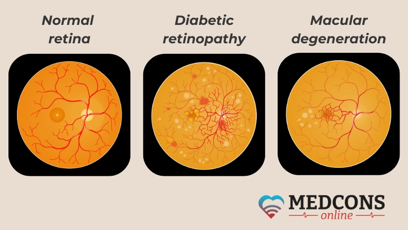
Age-related macular degeneration is one of the leading causes of blindness among the elderly. This disease consists in impaired central vision, which makes reading, driving and recognizing faces extremely difficult. The wet form of AMD is associated with abnormal growth of blood vessels that sprout under the retina and cause it to swell and haemorrhage.
Clinical trials have shown that anti-VEGF drugs such as ranibizumab and aflibercept significantly slow the progression of the wet form of degeneration. More than 90 per cent of participants had their vision preserved, and in a third of patients it improved by three or more lines on a standard chart. Aflibercept and other modern epithelial growth blockers act for a long time, which makes it possible to reduce the number of injections with a similar effect.
Diabetic retinopathy
This is a most severe diabetes complications threatening with complete blindness. It is caused by damage to small retinal vessels, which leads to haemorrhage, swelling and scar tissue formation. In severe cases, diabetic macular edema develops, with fluid accumulating in the macula, the central part of the retina responsible for visual clarity.
Anti-VEGF drugs show high efficacy in treating this dangerous disorder. According to studies, more than 50% of patients who received aflibercept and ranibizumab injections saw better vision, so these drugs are considered the gold standard for the treatment of diabetic retinopathy.
Central retinal vein occlusion
As a result of vein occlusion, the blood flow is blocked, causing poor blood supply, swelling and haemorrhages. This may happen suddenly, quickly leading to visual impairment, especially if the central veins have been affected.
Again, injections of substances that stop epithelial growth can significantly improve the situation by reducing the swelling, enhancing the blood circulation and restoring visual function. In clinical trials, more than 60% of patients reported an improvement in visual acuity after several months of treatment with aflibercept and ranibizumab.
Vabysmo: a new approach to treating retinal diseases
Vabysmo (faricimab) is an innovative bispecific antibody used to treat wet AMD, diabetic macular oedema and macular oedema due to retinal vein occlusion. The drug was developed by Roche and has received FDA approval in the US as well as approval from the European Commission.
It simultaneously blocks two factors that play a key role in the development of retinal diseases, i.e. VEGF and Ang-2. VEGF stimulates the growth of abnormal vessels, while Ang-2 destabilizes their walls, making them permeable. By blocking both of these factors, faricimab ensures more effective suppression of pathological processes in the retina.
Clinical trials of the drug showed its high efficacy in the treatment of AMD and diabetic macular oedema. In terms of vision improvement, it was comparable to the results of aflibercept treatment. However, the key advantage of the novelty is the possibility of longer intervals between injections. After the initial phase of treatment (4-6 monthly procedures), patients can receive Vabysmo every 3-4 months, while aflibercept is usually injected monthly or every two months.
Reducing the frequency of treatments significantly reduces patient burden and improves the quality of life. In addition, the medication has demonstrated a good safety profile in clinical trials.
In August 2024, Vabysmo received approval from the European Commission for the treatment of macular oedema associated with vein occlusion. This expands the drug's application and makes it a promising option for patients with various retinal conditions.
How the treatment is carried out
The efficacy of anti-VEGF therapy depends largely on the regularity of injections and careful monitoring of the retina.
Intravitreal injection procedure
The injections are performed on an outpatient basis and take a relatively short time. The procedure involves several important steps:
- Preparation: A thorough disinfection of the eye area with an antiseptic to minimize the risk of infection. A local anaesthetic in the form of drops is applied to the eyeball to reduce discomfort.
- Fixation and positioning: The doctor fixes the eye with a special eyelid dilator to prevent blinking and ensure accurate injection of the medication.
- Injection: The anti-VEGF drug is injected with a thin needle directly into the vitreous, a clear gel substance that fills the back chamber of the eye. The doctor makes a puncture through the sclera (fibrous membrane of the eye) a few millimetres from the cornea. The procedure takes only a few seconds and most patients do not experience significant pain, although there may be a slight sensation of pressure in the eye.
- After the procedure: It is recommended that the patients should sit with their eyes closed for a few minutes. Sometimes the doctor may prescribe antibiotic drops for a few days to prevent infectious complications. It is advisable to avoid eye rubbing and strenuous physical activity for the first 24 hours after the procedure.
Intravitreal injections are considered a safe procedure, but proper follow-up care and close monitoring for possible side effects are important.
Administration and dynamic monitoring regimens
There are several prescription options depending on the type of disease, its severity, and the patient's response to treatment:
- Monthly therapy. A series of injections are given at 4-week intervals for the first three months (start-up phase). This is the most intensive regimen aimed at maximum stabilization of the disease, especially in the initial stages. After three treatments, the doctor can assess their effectiveness and decide on a further injection regime.
- Prolonged therapy (Treat-and-Extend). This approach involves gradually increasing the intervals between injections depending on the condition of the patient's retina. After achieving a stable effect in the absence of relapse, the interval can be increased up to 6-8 weeks.
- On-demand therapy (PRN, from the Latin ‘pro re nata’). In this mode, injections are performed only when necessary, i.e., if the patient shows signs of relapse, such as visual deterioration or retinal swelling confirmed by optical coherence tomography (OCT). This format reduces the frequency of injections, but requires regular monitoring of the eye and frequent visits to the doctor.
Optical coherence tomography is the most important tool for dynamic monitoring. It is a non-invasive imaging tool that allows a detailed examination of the retinal structure and the detection of swelling or changes in the vascular system of the eye.
In addition to OCT, follow-up includes visual acuity testing and ocular fundus examination. As a rule, after the initial course of monthly injections, patients should be examined every 4-8 weeks, which allows timely detection of deterioration and adjustment of therapy.
Possible complications and side effects
Anti-VEGF therapy is a relatively safe method, but like any medical procedure, it is not without the risk of side effects and complications. The most common local side effects include:
- eye inflammation or irritation: short-time redness and mild pain after the injection;
- haemorrhage on the surface of the eye (subconjunctival haemorrhage): a relatively harmless condition that can look scary but usually goes away on its own within a few days.
Serious complications such as endophthalmitis (infection inside the eye) or retinal detachment are extremely rare, occurring in less than 1% of cases. These conditions require immediate care to avoid permanent damage to the ocular structure.
In general, a systemic side effect of anti-VEGF agents is an increased risk of cardiovascular events such as stroke or heart attack, especially in patients with comorbidities. However, in ophthalmology, such risks are minimal, as the drugs are administered locally and in low doses.
Conclusion
With the advent of anti-VEGF therapy, patients suffering from retinal diseases now have a chance to preserve their ability to see and improve their quality of life. Modern drugs such as ranibizumab, aflibercept and brolucizumab have proven effective in controlling disease progression and restoring visual function.
Despite the high cost of treatment and the need for regular injections, the use of medications that inhibit epithelial growth has been a revolutionary step in the treatment of retinopathies, changing the prognosis for many patients. In the future, with the development of new drugs and technologies, anti-VEGF therapy may become even more successful and affordable, offering people new hope for preserving their eyesight for years to come.
References
1. Arrigo, A.; Bandello, F. Molecular Features of Classic Retinal Drugs, Retinal Therapeutic Targets and Emerging Treatments. Pharmaceutics 2021, 13, 1102.
2. Khanani AM (2022) Jahrestagung der American Society of Retina Specialists (ASRS), New York City, USA, 13.–16.07.2022
3. V. Kakkassery, S. Winterhalter, A. M. Joussen. Die Rolle der Anti-VEGF-Therapie in der Behandlung des diabetischen Makulaödems. Verlag Unser Wissen (24. Mai 2021). ISBN-10:620372257X ISBN-13:978-6203722574
4. Schaal, Karen & Engler, Christoph & Schütt, F. & Scheuerle, Alexander & Dithmar, S.. (2008). Intravitreale Anti-VEGF-Therapie mit Bevacizumab bei neovaskulärer AMD. Der Ophthalmologe. 105. 538-543. 10.1007/s00347-007-1638-2.

Comments — 0