
Despite the fact that the mammary glands are easily accessible to external examination, the number of clinical errors in making a diagnosis is rather high. The error rate of standard mammography averages 38%, which makes breast conditions an urgent world problem. The main task of modern mammologists is to detect abnormalities at their early stages of development and recommend the most effective and sparing treatment.
What is digital mammography and how it works
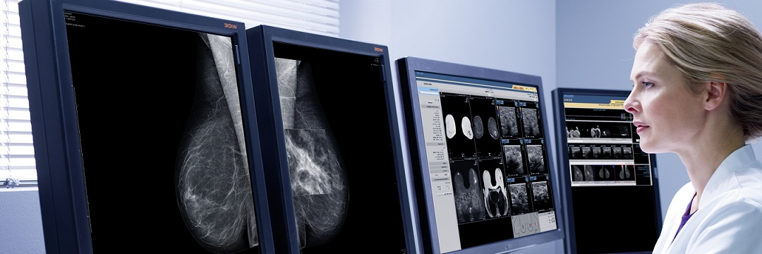
Digital mammography has replaced analog mammography. These units have a lower radiation exposure and greater sensitivity. A digital mammography machine is more compact in size than a standard one.
The X-ray tubes of a digital and standard mammography unit have virtually no difference. The difference is in the image recording. After the picture is taken, it is displayed as a digital image on the monitor screen.
When performing a digital mammography, X-rays pass through the breast and, with the help of detectors, transform the image into electronic impulses, which are transmitted to a computer digital matrix. With the help of special software, the image is converted into a clear visualization.
Advantages of the technique
Digital mammography is the gold standard for breast diagnosis. It has a number of advantages over standard units:
The image is projected on a monitor screen, stored digitally, printed out if necessary, and transmitted through an intra-hospital network.
The high sensitivity of digital mammography is in its capability of visualizing lesions smaller than 1mm.
If necessary, it is possible to enlarge the area of the picture requiring a detailed examination.
Computer programs allow one to change the imaging quality in order to improve the visualization of dense structures. It is possible to change the final version, convert the negative to positive, and create a scaled image.
The high sensitivity of the DR detector (a device for performing images without film) in digital mammography allows reducing radiation exposure by approximately 30%.
No x-ray films.
Digital mammography eliminates the need for duplicate films.
What makes mammography different from X-ray, ultrasound scanning and MRI
A mammography and an X-ray examination basically refer to the same examination method. The principle of their work is the use of X-rays. The only difference is the radiation dose. In mammography it is less than in X-ray exams.
Ultrasound examination is applicable for young women up to 40 years of age, since during this period, the mammary glands have a dense structure, and ultrasound allows to visualize even small changes. The technique is also used to monitor the condition of regional lymph nodes.
What are the ultrasound scan advantages?
Ultrasound is a safe and noninvasive technique for breast examination. Unlike mammography, there is no radiation exposure and no X-rays are used.
Ultrasound diagnosis can be used in women during pregnancy and lactation. It allows to determine diffuse mastopathies, abnormal lumps in the mammary glands more than 1.5 cm in size. Ultrasound technique is recommended for dynamic monitoring.
The examination is to be done on days 5-12 of the menstrual cycle, when the hormone levels decrease. This helps to avoid overdiagnosis or false positives. The ultrasound examination determines areas of the glands with increased and decreased echogenicity, evaluates the nipples, ducts and axillary lymph nodes. Currently, with the help of this tool, it is possible to diagnose cystic overgrowths in the breast with an accuracy of 93-100%.
Cooper's ligaments, which hold the breast and the pectoral muscle together, are usually poorly visualized. Ultrasonography has a lower informative value in fatty breast involution as well.
MRI
Magnetic resonance imaging is one of the methods of breast scanning. Most often it is recommended as an additional examination to clarify the diagnosis, or if ultrasound and mammography were not informative enough. The study does not involve the use of X-rays or other ionizing radiation.
There are two basic MRI techniques: with and without contrast. The application of contrasting agents increases the diagnostic value, opens up wider possibilities in comparison with X-ray and ultrasound examinations.
MRI allows to study abnormal findings in more detail, to reveal cancerous and benign neoplasms, diffuse and post-traumatic changes in the mammary glands. By means of modern devices it is possible to determine metabolic changes, conduct the spectroscopy of suspicious areas, visualize increased levels of blood supply, the latter usually indicating a malignant process. MRI allows detecting cancerous tumor degeneration at early stages of its development.
Second opinion
The digitalized mammography results enable remote consultations by high-level specialists. Today, every patient can seek a second expert opinion from doctors at the world's leading medical centers in order to get a fresh perspective on their issue, to find a more advanced or an alternative diagnostic and treatment option.
Comments — 2
Кристина
Я читала, что во время кормления грудью можно делать маммографию. Но ведь это же рентгеновские лучи. Действительно ли они безвредны?
Marina Virko
Современные аппараты цифровой маммографии являются очень щадящими с точки зрения рентгеновского излучения и безопасны для лактации. Для получения лучшего изображения в период кормления рекомендуется непосредственно перед началом обследования покормить ребенка или сцедить молоко. Это способствует снижению плотности молочных желез и, как следствие, повышает качество оценки снимков.