
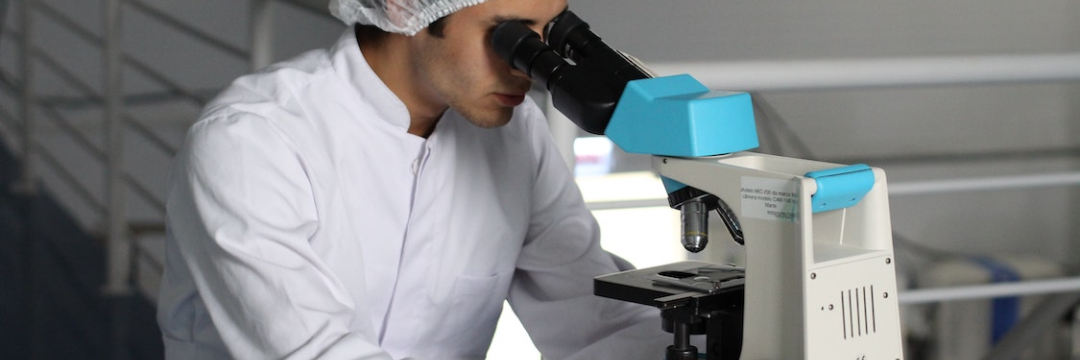
A study by US dermatologists has confirmed that it is worth taking another look into the microscope before a surgeon takes up the scalpel. Researchers from the University of California analyzed how often a diagnosis of a malignant skin tumor survived the scrutiny of expert dermatopathologists, and what impact the difference between the initial and second opinion had on the planned surgical procedure. At the University of California, it is a standard procedure for biopsies of patients in need of skin surgery to be re-evaluated by staff dermatopathologists before operation.
In the retrospective study, researchers compared the histological study findings of 358 patients referred by outside pathologists to confirm malignancy between January and December 2019 with assessments by the clinic's own experts. Patients who were first referred with a request for re-examination were excluded from the study.
In 10.3% of dermatology reports, the initial diagnosis of the external expert did not match the assessment of the UC examiner. In 31.6% of biopsies, re-examination revealed a different tumor subtype.
In 8.9% of cases, review led to a change in therapy. In the majority of cases the second opinion biopsy stated a less severe disease so that surgery could be avoided (87.5 %). 59.4 % of these cases were misinterpreted by the primary examiner as squamous cell carcinomas, 12.5 % as basal cell carcinomas. 15.6 % turned out to be melanocytic lesions on re-evaluation.
Only three of the 32 conflicting second opinion diagnoses revealed more severe findings than on external evaluation, and hence wider incision margins were necessary. Thus, on follow-up examination, one atypical nevus turned out to be melanoma in situ (MIS), and one MIS and one squamous cell carcinoma turned out to be invasive malignant melanoma.
In 79.9% of all biopsies, the initial diagnosis was made by an outside dermatopathologist, in 22.1% by a dermatologist without dermatopathological training, and in 5.6% by a pathologist. More often than not, assessments by dermatologists without dermatopathological training differed from the second opinion of experts (22.5% vs. 15.0% for pathologists and 8.4% for dermatologists with relevant qualifications).
References
https://www.jaad.org/article/S0190-9622(20)33178-9/fulltext
Comments — 2
Ольга
У меня на коже появилась крупная опухоль, ее удалили, при этом дерматолог сомневался, что опухоль похожа на злокачественную, но лаборатория в Харькове поставила диагноз: злокачественная меланома. Я отправила стёкла в Киев, там ответили, что опухоль скорее всего доброкачественная. Могу я прислать стёкла для новой оценки в Германию?
Marina Virko
Здравствуйте. Да, повторная оценка возможна. Вы также можете приехать и привезти с собой стекла и блоки для повторной экспертизы. Их надо обязательно исследовать для исключения ошибки, которая может иметь очень серьезные последствия. Также следует предполагать, что материал, прошедший через 2 лаборатории, может оказаться неполным. В том числе по этой причине в спорных случаях настоятельно рекомендуется действовать как при диагнозе меланома, то есть провести повторную резекцию в объеме 1 см от края удаленной опухоли. Также рекомендации по клиническому наблюдению области удаленной опухоли совпадают с послеоперационным наблюдением меланомы.