Digital mammography with tomosynthesis
Advantages of mammography with tomosynthesis
During the study, images are taken in the form of tomograms with a specified slice size. The results are digitized and provided in a convenient format for visualization.
The examination helps in the diagnosis of abnormal processes, which cannot be detected by a planar examination. Tomosynthesis allows to avoid false positive results, which are possible with other methods of examination. Its main advantages are:
- High accuracy compared to conventional mammography (more than 30%).
- Less radiation exposure.
- Primary abnormal changes in the breast are diagnosed 27-30% more frequently compared to other methods.
- High image quality.
- Low percentage of diagnostic errors, false positive results in regard to breast cancer.
- Ability to detect microcalcifications, cancerous formations at the earliest stages, and to differentiate cystic fibrosis mastopathy.
Once the images are obtained, they can be further processed using computer software, in order to improve their quality and get a clearer image.
Indications for mammography with tomosynthesis
Tomosynthesis (3-D mammography) is indicated for women who are at risk due to:
- A cancer history – when the patient anamnesis includes any malignant condition.
- A first-line family history of cancer.
- Injury, reconstructive breast surgery, including cosmetic procedures.
- Prolonged hormone replacement therapy (amenorrhea, menopause).
- Detected precancerous conditions (inflammatory and benign).
Contraindications and limitations
The examination, despite its effectiveness and safety, has a number of contraindications:
- Young women, under the age of 30, are not eligible for mammography with tomosynthesis due to increased breast density.
- Pregnancy and breastfeeding are also on the list of contraindications, due to the minor but existing radiation exposure.
- The presence of implants is a relative contraindication; they can complicate the diagnostic process.
Preparation for the exam
It is recommended to undergo the examination while estrogen levels are low, i.e., on days 5-12 of a menstrual cycle. In case of an emergency, mammography is performed regardless of the cycle. For menopausal women, the examination can be performed at any convenient time.
The procedure does not require any special preparation. If you have the results of additional diagnostic methods, you must bring them to your mammologist for review. Immediately before the procedure, it is not recommended to apply creams, gels, and deodorants to the area under the arm. All metal jewelry and details of clothing should be removed.
Duration of the entire procedure is 10 minutes on average, time of examination of one projection is 3-4 seconds.
Course of the examination
Before the examination, the patients are asked to remove her clothes to the waist. The mammary gland is placed on the plate, a clamp will press it from above. In the course of the procedure there is no need to compress the breasts too hard, thus the examination feels more comfortable as compared to the classical method. This is why tomosynthesis can also be performed in women with breast implants.
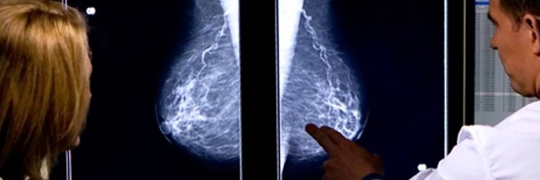
In contrast to conventional planar mammography, images are taken in two perpendicular views. By taking a series of pictures, it is possible to obtain detailed images of all abnormal areas. The collected data is transmitted as an image to the screen. 3D images can be created with the help of computer software.
Reading the results
Specialists need a little more time to interpret tomosynthesis findings as compared to conventional mammography. The results of the examination are saved on digital media and given to the patient on a disc. If necessary, the images are printed out on special paper using a printer.
Second Opinion
References
- Ingrid Mühlhauser, Birgit Höldke: Mammographie. Brustkrebs-Früherkennungs-Untersuchung. Kirchheim-Verlag, Mainz 2000
- H. Jung: Mammographie und Strahlenrisiko. In: RöFo: Fortschritte auf dem Gebiet der Röntgenstrahlen und bildgebenden Verfahren. 169, Nr. 4, 1998.
Comments — 2
Татьяна
Интересно, а насколько цифровая томография точнее обычной?
Oleg Gluz
Здравствуйте. Цифровая томография точнее обычной как минимум в несколько раз – за счет того, что рентгеновские трубки передвигаются вдоль поверхностных плоскостей молочных желез и производят съемку большего количества слоев ткани в большем количестве проекций.