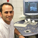
Any change in the female breast causes anxiety. But remember, there are many other conditions besides breast cancer that are much more likely to occur, such as a cyst. It is something like a small bubble filled with fluid, which is usually quite harmless. Nevertheless, a visit to the doctor is necessary to rule out cancer and plan further action.
What are cystic masses in the breast?
The female breast has about ten to twenty branched mammary glands (glandular lobules) of different sizes surrounded by connective and supporting tissue. Their function is to produce milk for feeding the baby, which is transported to the nipple through the milky duct system. But even without pregnancy, the breasts produce secretions. If the duct that drains the fluid is blocked, a cyst forms.
The size can vary from very tiny to several centimeters in diameter, and the cyst usually feels like an elastic ball to the touch. It typically occurs between 30 and 50 years of age. Statistically, this is the most common benign breast mass, which is detected in almost every forty-year-old woman.
What factors lead to breast cysts?
The causes can be various. For example, cystic changes are stimulated by an imbalance between estrogen and progesterone, i.e. a change in the hormone profile. Therefore, cysts occur in mastopathy, a hormone-dependent benign breast condition characterized by increased growth of glandular cells, resulting in the production of more fluid. Besides, the size and number of cysts can vary during the menstrual cycle.
It may also be associated with the following situations:
trauma to the breast tissue caused by its chronic inflammation, which leads to damage to the milky ducts and the small vessels through which fluid is drained;
damage to the fatty tissue, for example as a result of surgery or an accident;
congenital predisposition.
Symptoms: how does a cyst in the breast manifest itself?
In most cases, the fluid-filled masses do not cause any complaints and are discovered accidentally during palpation as a round, movable lump. In some women, the area surrounding the cyst becomes sensitive and painful, especially before menstruation. But when it reaches a certain size, it begins to press on the nearby tissue and cause pain. Unusual discharge from the nipple can also indicate the presence of a cyst.
When should I see a doctor?
Any self-detected changes in the breast are a solid reason for visiting a care provider, and even more so if there are pains or drops of liquid from the nipple. Particular attention should be paid to the following alarm signals:
- sudden changes: a noticeable change in the size or shape of an already known cyst;
- severe and persistent pain in the breast area;
- nipple discharge, especially if it is bloody or formed without squeezing;
- changes in the skin: redness, swelling or dimpling.
What is the diagnostic process?
The first step is a detailed interview, known as an anamnesis, in which the doctor finds out about circumstances that may be important for the development of further strategy, e.g.:
- The nature of the complaints, their duration and intensity.
- A relationship between the symptoms and the menstrual cycle.
- Chronic diseases.
- Family history: whether there has been a history of malignant diseases (especially breast cancer) in the immediate family.
- Regularly taken medications.
Then the doctor performs a thorough palpation of both breasts to identify lumps and check if they are movable. After that, additional workup is suggested to clarify the palpation findings.
Ultrasound
The test is based on the use of harmless sound waves. They are emitted by a special transducer and reflected by the tissues to varying degrees depending on their density. In this case, the fluid appears on the image as a darkening, and a cystic formation can thus be easily distinguished from a solid one (tumor). However, sometimes the contents can be viscous (if they contain blood components, protein flakes or pus), which makes differentiation difficult.
Ultrasound also makes it possible to determine the location and size of the cyst. In addition, an experienced ultrasound technician is able to assess its structure, recognize possible changes and growths on the inner wall, where papillomas and, in very rare cases, carcinomas can form.
Mammography (breast X-ray)
X-ray examination in the diagnosis of cystic changes is indicated in case of ambiguous ultrasound results or in older patients. It also shows other abnormalities such as calcifications, precancerous or malignant conditions.
Breast MRI
Magnetic resonance imaging may be performed at the patient's request but, in terms of clinical guidelines, is not considered necessary.
Fine-needle aspiration
When the cyst reaches a certain size (1.5 cm), to confirm the diagnosis, it is usually punctured, i.e. its contents are aspirated with a hollow needle and examined under a microscope for cellular changes. Nowadays, this type of biopsy is usually performed under ultrasound guidance.
The likelihood of papilloma and especially carcinoma formation on cysts is generally extremely low. However, if malignancy is detected, the cyst and surrounding tissue should be removed.
Classification of breast cysts
An important characteristic is the structure.
Simple cysts are filled only with transparent fluid, have clear boundaries, smooth walls with the absence of any inner structures. As a rule, they are completely harmless and do not require any action.
Complex cysts are characterized by the presence of internal partitions ordebris, the fluid may be opaque. Due to the internal heterogeneity, a tumor may be suspected, so additional diagnosis is recommended.
In terms of size, a distinction is made between microcysts (tiny masses that can only be detected by ultrasound, mammography or even a microscope) and macrocysts (2.5 to 5 cm in diameter, detectable by palpation).
Does a cyst in the breast need to be treated?
Therapy is not necessary if the mass:
- is small (less than 1 cm),
- is not growing,
- has smooth walls,
- has no internal partitions,
- and does not cause any complaints.
Large and painful cysts are punctured, removing the fluid content, which helps relieve pressure on the surrounding tissues. Once the cyst is emptied, it is usually filled with air, after which the empty cyst shell should be glued together with scar tissue. This measure prevents re-accumulation of fluid in the cyst.
The aspiration fluid is examined for malignant cells if it is cloudy, has an admixture of blood, there is little of it, or when the cyst persists after the procedure.
In other cases, a follow-up examination is carried out after 4-8 weeks. If by this time the cyst is no longer palpable, it is considered benign, and this completes the treatment. Otherwise, it is drained again with subsequent examination of the punctate.
Surgical removal is advisable for very large or recurrent cysts, as well in as for potentially malignant changes (e.g. microcalcifications). Complex cysts, which have a higher risk of developing carcinoma, are surgically removed in almost all cases. The operation is usually performed under local or general anaesthesia, depending on the size and location of the mass. The procedure can be carried out either through a small incision or by vacuum punch biopsy.
Prognosis and prevention
There is no effective prevention of cyst formation. The only preventive measure is self-monitoring of the breast. Inspections should be carried out monthly on the same day of the cycle. In case of suspicious findings, you should immediately contact a breast care specialist.
The prognosis is favorable. On the whole, the presence of these benign formations does not pose a threat to health and does not bring much trouble, so when they are detected, a wait-and-watch approach is usually chosen. The most important thing is to be sure that the nature of the lump has been determined correctly, that is, it is indeed a generally harmless fluid-filled cavity and not a tumor.
What should I do if I suspect a breast cyst?
The most important thing is not to panic. Bear in mind that in the vast majority of cases such issues are not about cancer. But indulging in carelessness is not good either. Neither does it make sense to waste time anxiously looking for answers in Internet forums or asking acquaintances. Just make an appointment with a doctor! Which one?
In the latter case, do remember about subspecialities, i.e. it is better to go to someone who is constantly engaged in breast diagnostics.
Second Opinion
The frequency and relative safety of cystic breast masses do not mean that, having learned about this diagnosis, you can relax. First, it is worth while making sure that it is true. If the node is very small, distinguishing a fluid-containing formation from a solid one is not always easy, whereas the difference is fundamental. Therefore, it is quite a reasonable practice to get a review of your imaging data.
Second, a proper disease monitoring plan is very important in this condition. Which tests are more informative in each specific case, and when they should be performed, depends on individual parameters - age, gland tissue density, hormone levels. The goal is not to overlook changes in the cyst itself or nearby tissues, which can lead to a more serious disorder. Therefore, if your doctor does not provide a detailed plan for some reason, or does not explain the essence and purpose of diagnostic procedures, you can seek a second opinion from another specialist.
When it comes to surgical treatment of a cyst, an independent evaluation by an experienced breast care provider is a way to make certain that this option is indeed unavoidable and the optimal one.
And one more (by no means the least important) argument: it always makes sense to have as much understanding of your disease as possible, including the way it can develop, its treatment options and prognosis. A primary care physician does not always have the time or ability to provide such details. However, you can get them from world-class experts using the breast care second opinion service.
References:
- Breast Cyst Fluid Analysis Correlations with Speed of Sound Using Transmission Ultrasound
- Breast cyst aspiration. Can Fam Physician. 2012
- Breast Cyst. 2021.
- Human gross cyst breast disease and cystic fluid. 2006.


Comments — 1
Комаровская Евгения
У меня кисты в груди примерно с 30 лет. Когда я в первый раз нащупала кисту, а это было в третьем триместре беременности, сразу помчалась к врачу. Врач решила перестраховаться и отправила меня маммографию. Хорошо, что рентгенолог отказался и еще раз сделал УЗИ. С тех пор я постонно делаю и УЗИ, и маммографию, недавно выявили крупную кисту, ее на всякий случай пунктировали, оказалась доброкачественная.