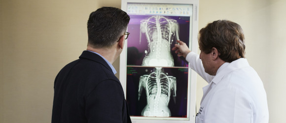
Expert opinion and online consultations available on your computer or any mobile device. The service is available wherever there is an internet. All you need is a PC or a smartphone.
Malignant tumors of the testes are not widely spread in general. But it is the most common cancer in men between 20 and 40 years of age. The diagnosis sounds frightening; however, the good news is, it can be cured. The outcome largely depends on correct treatment strategy. A second opinion on testicular cancer is therefore highly recommended to ensure the best results.
Origin and types
In more than 90 percent of cases, malignant growth in the testes arises from male reproductive cells (the so-called germ cells); accordingly, the majority of such masses are germ cell tumors. The remaining 10% develop from supportive tissue (stromal cells) and become malignant only very rarely.
Germs cell tumors are further divided into seminomas (slow-growing type) and non-seminomas (that are more aggressive).
Exact causes of testicular cancer have not been defined, but men with undescended testicles, even after correction in childhood, and men with relatives who were affected (e.g., father or brother) have a definitely higher risk.
Symptoms
In its early stages, the condition does not cause any symptoms. With time, patients may notice a lump or swelling in either testicle and an increase in its size. Other possible signs include a feeling of heaviness in the scrotum, pain or discomfort, as well as enlargement or tenderness of the breast tissue.
Diagnosis
A tentative diagnosis can usually be made at a clinical examination. The next step will be an ultrasound scan of the testicles and the abdominal cavity, which provides essential data for further decisions. If a mass is detected, an exploratory surgical procedure will follow. Should it confirm a tumor, the affected testicle together with the spermatic cord is removed. A histological examination of the removed tissue is then performed to determine the exact type of the disease (seminoma or non-seminoma).
Imaging studies, such as computed tomography of the abdominal cavity and thorax, are made to evaluate the tumor stage (whether and how far it has spread into other areas).
Blood tests aimed at looking for specific tumor markers (proteins which are released by testicular tumors in varying amounts depending on their type and stage) give significant information both in terms of primary diagnostics and follow-up case monitoring.
Staging and histology
As in the case of other malignant conditions, tumors of the testes are classified into stages according to their extent. The key criteria are, in particular, the involvement of lymph nodes in the abdominal cavity (retroperitoneum) and metastases growth in other organs, usually in the lungs and liver:
stage I: no evidence of metastasis;
stage II: retroperitoneal lymph node metastases;
stage III: affected lymph nodes in the thorax (mediastinum) or distant metastases.
Testicular cancers differ in their cell structure. The majority of them are germ cell tumors which fall into seminomas and non-seminomas. The former may be classic or spermatocytic (occurring more frequently in older men and having an excellent prognosis). The latter are very variable and most more than one type is present in one patient. The four main non-seminoma types are: embryonal carcinoma, yolk sack carcinoma, choriocarcinoma, and teratoma.
Histologic features are of major importance since they determine the way a tumor will react to a particular treatment, as well as overall disease prognosis.
Treatment
Surgery is usually the primary option, which includes the removal of the affected testicle, and, in some cases, a biopsy of the other testicle in order to rule out simultaneous malignancy. This procedure does not affect the reproductive function, as the healthy counter-testicle takes over the function of the ill one. The follow-up treatment depends on the tumor histology, the stage of the disease, as well as tumor marker values.
Further operations may sometimes become necessary. In particular, if exams prove that the cancer can spread, or has already spread, to the lymph nodes, retroperitoneal lymph node dissection (RPLND) is recommended. It involves resection of lymph nodes in the posterior abdominal cavity. In most cases, the procedure can be performed in a nerve-sparing manner, so that the ability to ejaculate is preserved.
In certain cases, tumor manifestations (metastases) to other body parts can also be treated surgically. Depending on the testicular cancer type and stage, other treatment options will include systemic chemotherapy and, in the case of seminomas, radiation. Often a combination of therapies is recommended.
A second opinion matters
Despite the availability of evidence-based guidelines, determining the optimal therapy for testicular cancer is obviously a challenge. In particular, studies conducted by the German Cancer Society have shown that around 30 percent of prescribed therapies deviate from general recommendations. To avoid mistakes, patients are encouraged to seek second opinions from experts at specialized centers with extensive experience in research and clinical practice. For example, such advice is of special value when it comes to very complex surgical procedures to remove residual tumor tissue after chemotherapy.
What is the service about?
A second opinion on testicular cancer is a service which makes it possible to get a remote consultation of a qualified specialist, based on available medical summary or study results.
It might be helpful:
• to confirm the existing diagnosis;
• to make sure that the recommended treatment is correct;
• to obtain information on advanced methods of testicular cancer diagnostics and treatment;
• to get expert commentary on previously performed exam results;
• to make the right choice if there are two or more possible therapeutic options.
What will the client get?
A diagnostic conclusion, as well as recommendations on treatment and follow-up, based on the provided information. If the initial data is incomplete, further examinations will be suggested.
What data should be provided to get a second opinion?
Written reports:
Attending physician’s statement (preferable)
Scrotum ultrasound (mandatory)
Histopathology (mandatory, if it was performed)
Tumor marker laboratory tests (preferable)
Overall reports size: up to 5 pages
Imaging scans:
Thorax and abdomen CT (optional)
Overall imaging volume: up to 2 exams
What are the second opinion formats and terms?
Written second opinion:
Making a report based on the data provided, the consulting specialist summary including a diagnostic report and recommendations for further diagnostic, treatment and monitoring tactics. Report size: up to 1 page.
Video consultation:
All services of written second opinion. Additionally: a 10-minute video consultation with a doctor, including a visual patient examination, clarification of symptoms, radiology images consulting, explanation of the proposed treatment tactics, answering patient's questions.
Phone consultation:
All services of written second opinion. Additionally: a 10-minute telephone consultation with a doctor, including clarification of symptoms, explanation of the proposed treatment tactics, answering patient's questions.
Specialists in Testicular cancer
You do not have to spend hours getting through busy hospital lines, or sitting in waiting rooms. Expert advice will be delivered fast and free of your effort.
Sort by: [[ s.txt ]]sortsort
Nothing found, try changing search options
