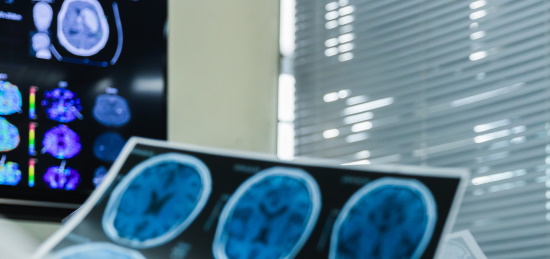
A re-evaluation of PET-CT scans by expert radiologists. The service is available wherever there is an internet. All you need is a PC or a smartphone.
PET CT Second Opinion
PET-CT combines two types of radiological diagnostics. Computed tomography produces high-resolution images of cross-sections of the body. Based on these, the radiologist assesses the anatomy of the body, identifies changes in the organs, and determines their nature. At the same time, positron emission tomography technology can be used to draw conclusions about certain metabolic processes in the tissues.
Before a PET study, the patient is injected intravenously with a radiopharmaceutical. This is a low-radioactive substance that is distributed throughout the body. It almost never causes allergic reactions. The natural decay of this substance produces low radiation, which allows the doctor to visualize its distribution in the body. With the help of computed tomography, which is performed simultaneously, these metabolic processes can be accurately attributed to specific parts of the body.
The most commonly used PET-CT radiotracer is 18F-FDG, a glucose analog with the positron-emitting radionuclide fluorine-18, which displays cell metabolism. Since many tumor cells have an increased metabolism (e.g., due to their rapid growth), the radiopharmaceutical allows them to be detected.
In other words, it is PET-CT that can simultaneously answer two fundamentally different but very important questions:
- "What does the tissue look like?"
- "What are its biochemical properties?"
It can help, for example, to assess the effectiveness of chemotherapy for malignant diseases. Very often, metabolic changes in tumor foci become visible on PET-CT long before the mass actually shrinks in size. Because chemotherapy disrupts the metabolism of tumor cells, they absorb less radioactively marked glucose. Therefore, the doctor can determine early enough whether chemotherapy is the right choice or whether the treatment plan needs to be changed.
PET-CT interpretation
The scan is only one step in the diagnostic process. The resulting images must be read by a physician. For this purpose, the functional positron emission scan data are combined with anatomical data from a CT scan. The images may show areas with increased metabolic activity, which may indicate the presence of tumor foci or inflammation. The metabolic activity assessment unit is SUV (standardized uptake value).
Computed tomography is used to determine the exact location and size of a lesion.
The final result of PET-CT image analysis is a report. It is prepared by a radiologist and includes a detailed description of anatomical and functional data, including localization and size of areas of pathological metabolic activity.
Why may I need to have my PET-CT findings reviewed?
The combination of PET and CT in the form of fused images plays an important role in the diagnosis and treatment of a number of complex diseases. Therefore, a patient's life often depends on how accurately the obtained data is interpreted.
A second opinion on PET-CT from a highly qualified radiology specialist may be required if:
there is no certainty about the diagnosis;
there are doubts that the initial reading of your imaging study was correct;
the available description of your PET-CT scan is not detailed enough;
your condition and/or issue requires an assessment by an expert with a certain subspecialty (e.g. cancer radiology or neuroradiology);
two or more therapeutic strategies can be considered as possible options.
What is a second medical opinion on PET-CT?
The result of the consultation is a written opinion by a radiologist on your PET-CT scans, with comments on the quality and significance of the available imaging data, a detailed reading of the scans, conclusions on the possible nature of any suspicious lesions, neoplasms or other abnormalities detected. The doctor's second opinion may also include an assessment of disease course (if previous data are provided for comparison) and recommendations on further strategy (such as further diagnosis of the detected abnormalities or regular follow-ups).
What records do I need to share to obtain a PET CT second opinion?
The following data is requested to obtain a reading and an assessment of radiologic study findings:
- files with current PET/CT images;
- files with the previous study (in case comparative evaluation is necessary);
- anamnesis (in the form of a doctor's report or a completed questionnaire).
What are the available PET-CT second opinion types?
Written second opinion:
- An expert radiologist’s report with a PET-CT scan readings and the specialist's conclusions. Report size: up to 1 page.
Video consultation:
- All services contained in the written second opinion plus an up to 15-minute video conference with the radiologist in which the consulting expert comments on the imaging, explains the conclusions, discusses the recommended plan and answers the patient's questions.
Phone consultation:
- All services contained in the written second opinion plus an up to 10-minute telephone talk with the radiologist in which the consulting expert explains the conclusions, discusses the recommended plan and answers the patient's questions.
Specialists in PET CT Second Opinion
You do not have to spend hours getting through busy hospital lines, or sitting in waiting rooms. Expert advice will be delivered fast and free of your effort.
Sort by: [[ s.txt ]]sortsort
Nothing found, try changing search options
